
Zymography: What Activity Bands Can Show About Lumbrokinase Samples
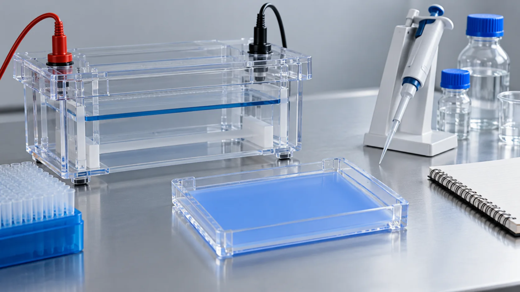
Zymography: What Activity Bands Can Show About Lumbrokinase Samples
Zymography separates proteins in a gel and reveals where an enzyme can degrade a chosen substrate under the assay conditions. For lumbrokinase samples, an activity band can support a characterization study, but it does not independently establish identity, purity, quantitative potency, or a health effect. The value of the image depends on the substrate, controls, sample preparation, and complete experimental record.
Buyers evaluating lumbrokinase powder should therefore ask what question a zymogram was designed to answer. A development laboratory may use it to investigate activity patterns across fractions or batches. Routine batch acceptance may rely on a different, agreed quantitative assay. These pieces of evidence can complement each other without being interchangeable.
How does a protease zymogram produce an activity band?
In a common substrate-gel approach, proteins migrate through a polyacrylamide gel that contains a material susceptible to proteolysis. After separation, the gel undergoes the recovery and development steps needed for the selected enzymes. Staining makes the remaining substrate visible, while local substrate degradation produces clearer regions.
Bio-Rad’s guide to protease analysis by zymogram PAGE describes this principle using gelatin or casein substrates. The guide separates electrophoresis, enzyme recovery, substrate digestion, and staining as distinct stages. Its product-specific procedure should not be transferred unchanged to an earthworm enzyme sample without checking suitability.
In Thermo Fisher’s electrophoresis handbook, the described gelatin-zymogram workflow uses nonreducing, unheated sample preparation followed by renaturation and development. That is an example of an established system, not a universal rule for every protease. The receiving laboratory should identify the preparation appropriate for its target activity.
For procurement, the useful distinction is between the presence of protein and the activity detected by this particular system. A conventional protein stain and an activity zymogram answer different questions even when their lanes contain samples from the same batch.
Why does the substrate name matter for lumbrokinase?
The substrate determines which degradation event the assay can reveal. Gelatin, casein, fibrinogen, and fibrin should not be treated as equivalent labels. A laboratory report should name the substrate and describe how it was incorporated or prepared before interpreting the clear zones.
A 2023 original study of enzymes extracted from Lumbricus rubellus used fibrinogen zymography alongside protein separation, fibrin-plate testing, and proteomic analysis. The researchers examined a particular experimental preparation and its fractions. Their results illustrate complementary characterization methods; they do not establish the band pattern, activity grade, or composition of a different supplier’s material.
For an inquiry, replace a vague request for an enzyme gel with a precise question: does the laboratory have a suitable method for detecting activity against the substrate relevant to this project? Ask how the positive control demonstrates that the complete method works.
What does published lumbrokinase research actually demonstrate about the method?
Published methods show that gel-based activity analysis has been used in lumbrokinase research. They do not show that every commercial powder has undergone that analysis or that an image from a paper represents a product being offered for sale.
For example, a 2010 study involving Eisenia andrei-derived lumbrokinase described a fibrin zymogram prepared with fibrinogen and thrombin, followed by detergent exchange and incubation. That paper studied experimental materials in an animal-model setting. Its laboratory method is relevant background, while its biological findings should not be presented as evidence of human absorption or a benefit from a supplier’s ingredient.
When reviewing a cited paper, keep a short record of the studied material, substrate, preparation, and purpose. Then ask which of those conditions match the commercial sample. Where there is no demonstrated match, retain the paper as research context rather than treating it as a batch certificate.
How is zymography different from other analytical evidence?
Zymography connects electrophoretic migration with detectable substrate degradation. Other methods provide different information. The right evidence set depends on whether the project is investigating identity, activity, consistency, or a change in a sample.
| Evidence | Question it can help address | What should not be inferred automatically |
|---|---|---|
| Substrate zymogram | Where is activity detectable under this gel method? | Complete identity, quantitative potency, or clinical effect |
| Protein-stained electrophoresis gel | What separated protein pattern is visible? | That every visible protein is active |
| Agreed quantitative activity assay | What activity is measured under a specified procedure? | That different assay units are interchangeable |
| Mass spectrometric characterization | What sequence-related evidence supports identification? | That identification alone establishes retained activity |
| Batch COA and sampling record | Which reported results belong to the shipment? | That a generic research image represents that batch |
Venacorebio’s current product information lists fibrin plate assay as the activity method. A request for zymography should therefore clarify whether it concerns additional characterization, rather than silently replacing the stated assay. Availability and scope of any extra testing need written confirmation.
What should buyers request with an activity-band image?
Request enough information to reconstruct the comparison. A cropped picture with unlabeled lanes cannot establish which sample produced which band. The report should identify the product, batch or fraction, extraction or dilution history, lane assignments, and loading basis.
Ask for the original image with the relevant surrounding lanes and controls. If a presentation image has been adjusted, the report should retain the unprocessed record and explain the adjustment. Compare samples using a consistent imaging approach rather than choosing different display settings for each lane.
The loading basis deserves particular attention. Equal volumes of two solutions do not necessarily contain equal amounts of protein or equal starting activity. State whether comparison is based on powder mass, measured protein, or a specified activity assay. Each approach asks a different question and should be interpreted accordingly.
Also request method identification and a clear conclusion from the responsible laboratory. Distinguish an observation such as a new clearing zone from an interpretation such as a changed activity profile. A purchasing team should be able to see which part is directly observed and which part needs further investigation.
Which controls make the comparison more informative?
Controls should address the specific risk of misinterpretation. The laboratory should explain what each control tests and which outcome would make the run unsuitable for comparison. The following is a discussion checklist for a characterization project, not a universal release protocol.
| Control or comparison | Question to discuss with the laboratory | Reason for including it |
|---|---|---|
| Appropriate positive control | Does it show that the selected substrate and development system can reveal the intended activity? | Helps distinguish sample findings from a failed assay system |
| Preparation blank | Does the extraction or dilution medium create unexpected background? | Checks a source of misleading clearing or staining |
| Reference sample | Is a retained, documented comparator run with the new sample? | Supports a controlled batch-to-batch comparison |
| Loading series | Are several suitable sample loads evaluated when signal intensity matters? | Helps identify overload and an appropriate comparison range |
| Independent preparations | Does the observed pattern recur after fresh sample preparation? | Checks whether the observation is reproducible |
A reference sample should have an agreed purpose and documented handling history. Calling it a reference does not make it an official standard or give it a certified potency. Keep that distinction clear when requesting a laboratory quotation or comparing reports.
Does a larger clear band mean higher potency?
Only a validated quantitative relationship could support that interpretation within its demonstrated range. Band appearance can depend on the sample load, development interval, substrate, recovery conditions, and imaging. A dramatic clear region by itself is not a potency unit.
The original 1997 study on quantifying activity in substrate gels investigated a specific staining method and established quantitative behavior for its experimental system. It illustrates why linear range and reproducibility need evidence. Its detection performance cannot be assigned to a different enzyme preparation or commercial powder without validation.
For a batch investigation, ask the laboratory whether the proposed image comparison is qualitative, semiquantitative, or quantitatively validated. Make sure the final report uses the same level of interpretation. If the project needs a numerical activity result, specify the assay and reporting units separately.
Do not convert band brightness into a capsule dose or a predicted biological effect. Those conclusions require evidence outside the gel experiment and fall outside an ingredient-characterization report.
What does a weak or missing band mean?
A missing band means the assay did not reveal a clear signal under the tested conditions. It does not, by itself, prove that the original powder contains no active enzyme. Begin by checking that the controls and sample-preparation records support interpretation of that run.
An established Methods in Enzymology discussion of protease zymography identifies recovery after SDS exposure and development conditions as important limitations. It also describes how prolonged development can reduce band resolution. These points explain why an unsuitable method can conceal or blur information rather than simply producing a weaker version of the correct answer.
Ask for a reasoned investigation instead of repeatedly increasing sample load until a visible zone appears. The laboratory may need to examine preparation, substrate suitability, inhibition, or development conditions. Record any changes so results generated by different procedures are not combined into a misleading trend.
Can band position identify a particular lumbrokinase component?
Band position can guide a characterization question, but position alone does not uniquely identify a protein. Report migration as an observation under the stated method. Any molecular-mass estimate should be appropriate to the gel system and its calibration, and should be distinguished from confirmed identity.
If a project requires identification of an active component, discuss a suitable combination of separation, activity detection, and independent identification. That may require coordinated sample preparation across different analyses. Agree on how the evidence will be connected before the sample is consumed.
More bands should not automatically be described as greater purity, and fewer bands should not automatically be described as better quality. Interpretation depends on the material being supplied and the purpose of the assay. For a complex preparation, the relevant question may be a reproducible profile rather than a visually simple pattern.
How can zymography contribute to a batch investigation?
Use it as a defined comparison within a broader investigation. State the trigger, such as an unexpected quantitative activity result or a changed characterization profile. Identify the samples to compare, the method to use, and the decisions that different findings would support.
Run a documented comparator where appropriate and keep the investigation connected to the same batch records used for the other tests. If the image changes while the agreed activity assay remains comparable, that is a question to investigate rather than an automatic rejection or approval.
Likewise, a familiar image should not override a confirmed failure of an agreed specification. Release decisions belong to the complete quality system and the applicable purchase agreement. The gel report should state its scope clearly enough that a reviewer cannot mistake supportive characterization for the entire release decision.
Before routine adoption, decide whether the test is technically suitable, reproducible, and useful for that material. An interesting research method is not automatically a practical incoming-control test. Sample requirements, turnaround, expertise, and the action triggered by a changed pattern all deserve review.
What belongs in a laboratory request?
Describe the sample as accurately as the available documents allow. Include product identity, batch or fraction, powder or solution form, storage history, and known excipients or preparation components. Explain whether the goal is substrate-specific activity detection, a comparative profile, or investigation of an unexpected result.
Request a written proposal naming the substrate, preparation approach, controls, loading basis, image deliverables, interpretation level, and any suitability work required. Ask which activities the laboratory can support instead of presuming that a commercial gel system is already validated for the sample.
Connect the request to the supplier file. A laboratory can generate useful experimental evidence, while the supplier remains responsible for identifying the material and explaining its documentation. Neither party should claim that a gel image proves authorization for a destination market or substantiates a finished supplement’s health claims.
FAQ
What does zymography show in a lumbrokinase sample?
It can show where substrate-degrading activity is detectable after electrophoretic separation under a specified method. The substrate, preparation, development conditions, and controls determine what that observation means. It is supportive characterization rather than a complete description of the ingredient.
Is fibrinogen zymography the same as fibrin zymography?
No. The substrate preparation differs, and the report should name it accurately. Ask what degradation event the assay measures and how the method suits the project. Do not relabel a gelatin, casein, or fibrinogen result as direct evidence from a fibrin-substrate assay.
Can a zymogram replace the COA activity result?
Not automatically. A routine quantitative result belongs to an agreed assay with its own method and units. A zymogram may contribute additional evidence, but substitution would require a justified method decision and an agreed quality arrangement.
Does no visible band mean the ingredient is inactive?
Not necessarily. First check controls, preparation, substrate suitability, and recovery conditions. Report the observation as no detectable signal under that method until the laboratory has enough evidence to make a broader conclusion.
Does a clear activity band demonstrate a human health benefit?
No. A gel experiment is laboratory evidence about substrate degradation. It does not establish absorption, clinical effectiveness, a safe dose, or a treatment claim for a commercial ingredient or finished product.
Send Your Ingredient Inquiry to Venacorebio
Share your target application, activity-assay requirements, sample identity, destination market, and requested characterization documents. State whether zymography is a research question or an additional quality investigation. Review Venacorebio’s ingredient supply information and use the form below to discuss available product documentation, sample terms, and testing scope.